
How Space Is Opening a Window Into the Brain
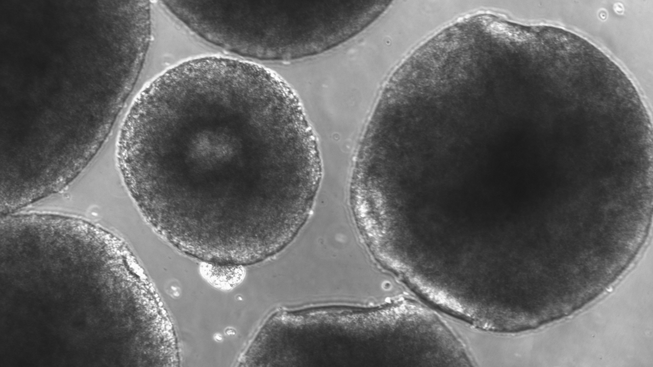
Human brain organoids grown in Alysson Muotri’s lab form three-dimensional clusters of neural cells, giving researchers a way to study brain development, disease, and the effects of spaceflight in a dish.
Media Credit: Alysson Muotri/UC San Diego
July 28, 2026 • By Stephenie Livingston, Senior Staff Writer
What came back from the International Space Station (ISSInternational Space Station) in 2019 was, to the naked eye, almost nothing. A few specks, roughly the size of a pea, suspended in liquid inside a sealed container. Under a microscope, though, they resolved into something stranger: tiny spheres of human cells that had, over months, organized themselves into rough three-dimensional models of human brain development. Grown without blood vessels or sensory input, they lacked consciousness, but had, after just 30 days in orbit, begun to show signs of aging.
When Alysson Muotri, a neuroscientist at the University of California, San Diego, ran molecular analyses on these brain organoids, he observed that the molecular clocks inside neurons, which scientists use to estimate biological age, had lurched forward by about a decade. It was as though the cells had lived through years rather than days.
“That was the eureka moment,” Muotri said. “I said, oh my gosh, this is senescence happening in accelerated time.”
Muotri’s finding suggested that spaceflight may accelerate molecular processes linked to neurodegenerative disease. That matters because the brain is hard to study in decline. Its neurons largely do not regenerate; they accumulate damage over a lifetime, making diseases like Alzheimer’s, Parkinson’s, and ALS slow and difficult to model. Animal studies often fall short, and brain organoids grown on Earth follow the same slow developmental clock as the organ they mimic.
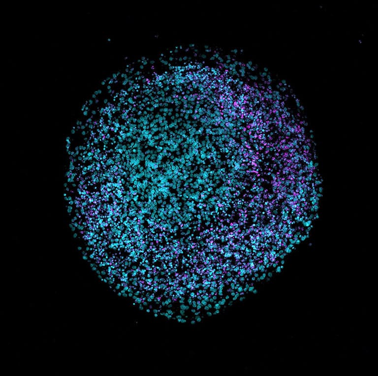
2D cryosection of a human brain organoid grown in a laboratory on Earth and stained with DAPI (teal) and VIPR2 (magenta).
Media Credit: Nreis1/Wikimedia Commons
Space may offer one shortcut, allowing researchers to see disease-relevant signals sooner. For patients and families watching symptoms advance, time matters. A faster model could help scientists see earlier what usually takes years to unfold and test interventions more quickly.
For Muotri, who came to this work through his son, those stakes are personal. His early career was focused on brain evolution and what separates humans from Neanderthals. Then he became the father of a child with severe autism. His academic questions led to a search for whether anything he found in the lab might translate into help for his son and others like him.
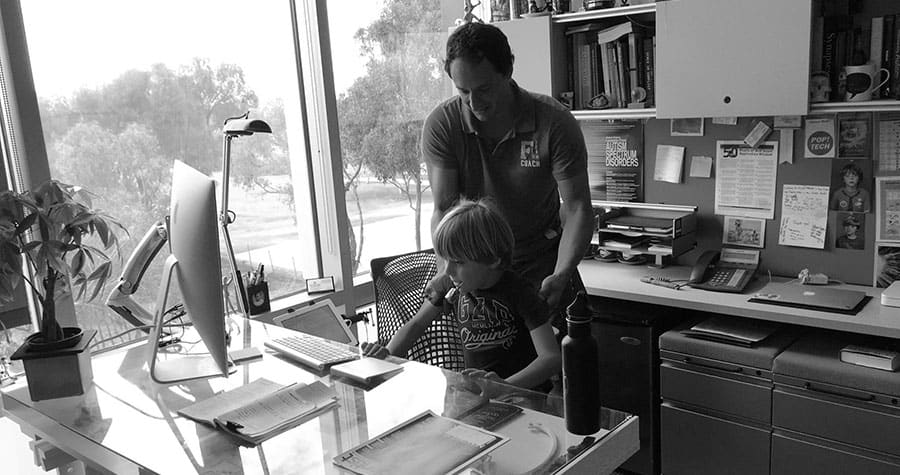
Alysson Muotri with his son, Ivan C. Muotri, who inspires his work with brain organoids.
Media Credit: Alysson Muotri
Brain organoids offered one path to explore those questions. Grown from induced pluripotent stem cells (iPSCs) derived from a patient’s own skin or blood, they self-organize into three-dimensional structures resembling regions of the human brain. They carry the donor’s own genetics, allowing scientists to study human brain biology in ways that animal models can’t replicate. Like any model, they are incomplete. They do not reproduce the full structure of the cerebral cortex, the vascular system that feeds the brain, or the billions of neurons and intricate circuitry that shape human thought and behavior. But models do not have to be perfect to be useful.
Most labs keep organoids for two months. A handful push to six months or a year. Muotri’s lab held the record at three years, until a researcher dropped the plate and contaminated the sample, which remains one of his more painful footnotes. “So, it’s not practical, right?” Muotri said.
Gravity normally drives convection in liquid, a constant mixing that disperses the signaling molecules and gases that cells use to communicate. Without it, those molecules accumulate differently around the cells, altering the chemical conversations neurons have with their neighbors. In space, something about that altered environment appears to speed things up.
Building a Brain Lab in Space
The ISS National Laboratory® has become a hub for neuroscience, connecting researchers with funding, flight opportunities, and the partners needed to move experiments from the laboratory bench to low Earth orbit(Abbreviation: LEO) The orbit around the Earth that extends up to an altitude of 2,000 km (1,200 miles) from Earth’s surface. The International Space Station’s orbit is in LEO, at an altitude of approximately 250 miles.. Michael Roberts, chief scientist for the ISS National Lab, said the field has evolved from a few exploratory studies into a growing body of evidence spread across multiple research teams and disease models.

Paula Grisanti, CEO and founding member of the National Stem Cell Foundation.
Media Credit: National Stem Cell Foundation
“What got our attention wasn’t a single result,” Roberts said. “It was seeing similar patterns emerge across different investigations led by different teams on different missions using different experimental designs and disease models.”
Paula Grisanti was one of the first researchers to test those ideas in space. The National Stem Cell Foundation (NSCF) CEO and co-founder was drawn into the possibilities of space research in 2017 during conversations with Space Tango co-founder Kris Kimel and the ISS National Lab team.
“I came out of that a total space nut,” she said. “I finally understood what it would mean to be able to watch cells behaving and interacting with each other in a way that wasn’t possible on Earth.”
ISS National Lab Commercial Service ProviderImplementation Partners that own and operate commercial facilities for the support of research on the ISS or are developing future facilities. Space Tango works with researchers to develop autonomous systems that keep organoids alive in orbit for weeks at a time, maintaining temperature, gas exchange, and pH without constant astronaut intervention.
Grisanti assembled a team, including stem cell biologists Valentina Fossati and Scott Noggle at the New York Stem Cell Foundation and Jeanne Loring at Scripps Research. They flew their first mission in 2019, the same year as Muotri, using what the team describes as regionalized neural organoids. These 3D human neural models begin as iPSCs in a 2D culture, where the cells are guided toward neural fates and toward specific developing brain regions. The team then transitions them to a 3D phase, where the cells continue to self-organize into more complex, tissue-like structures without further guidance.
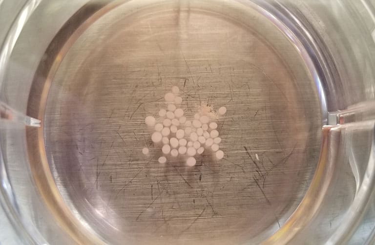
Brain organoids grown in Alysson Muotri’s lab cluster in a culture dish.
Media Credit: Alysson Muotri/UC San Diego
As the field has grown, researchers have moved toward more precise names for these brain cell-derived models because different labs build their systems differently, depending on the question they want to ask. Some models, like Muotri’s, are left to organize more broadly on their own, while others like Grisanti’s are guided toward particular brain-like regions so researchers can study specific processes. Those choices can shape what each model shows and may help explain why results from different model systems do not always align.
The NSCF team’s models matured faster in microgravityThe condition of perceived weightlessness created when an object is in free fall, for example when an object is in orbital motion. Microgravity alters many observable phenomena within the physical and life sciences, allowing scientists to study things in ways not possible on Earth. The International Space Station provides access to a persistent microgravity environment. than their earthbound counterparts, and their published results provided an early foundation for future work with brain organoids on the ISS. Their focus is on Parkinson’s disease and primary progressive multiple sclerosis, specifically on whether organoids derived from patient cells can reveal shared biology across conditions that appear clinically quite different. NSCF says it was the first group to send a patient-derived neural model that incorporates microglia, the brain’s immune cells, into space. Noggle, now a researcher with NSCF, suspects there’s an inflammatory pathway centered on microglia that underlies several neurodegenerative diseases.
“What we’ve come to understand over all of these missions is that there’s a common thread of things that happen in microgravity,” Noggle said.
He’ll also tell you they’re still working out what microgravity is doing.
Across six missions, overlapping gene networks keep appearing in the NSCF team’s system. Many are already implicated in Parkinson’s and Alzheimer’s disease, which Noggle sees as a sign that the changes aren’t random artifacts of spaceflight but reflections of biology that matter on Earth.
The next three ISS National Lab- and NASA-sponsored flights will add Alzheimer’s organoids to the mix, including cells carrying an early-onset mutation. Grisanti believes the work will lead to a drug-screening platform built on human brain tissue models, one that pharmaceutical companies and researchers could use to develop and test treatments years before disease would normally emerge.
But some of the most intriguing results have come from diseases that aren’t typically associated with aging at all.
A Disease Understood in Space
Rett syndrome occurs in young children after a period of apparent normalcy. Caused by a genetic mutation, the disorder primarily affects girls. A child learns to walk and use words, then something begins to unravel.
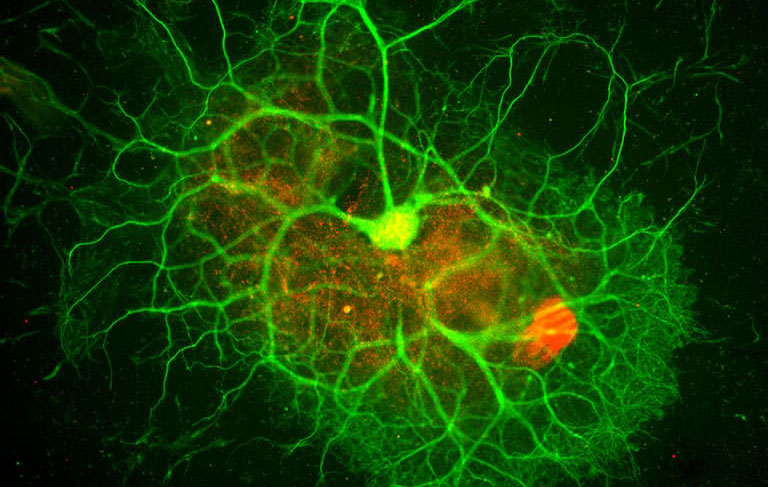
A brain organoid's human neurons branch into bright green networks.
Media Credit: Alysson
Once classified under the autism spectrum, it now carries its own diagnosis, marked by a brutal regression in the second year of life: language goes, then motor control, then the ability to eat. Muotri had modeled Rett in organoids for years, but his Earth-based models kept stalling right at that cliff just before the first symptoms appeared. That critical window, the moment the system crashes, sat exactly at the edge of what his models could show.
So, when Rett syndrome organoids returned from one of Muotri’s ISS missions showing senescence-associated and inflammatory signatures that had not appeared as clearly in his Earth-based models, they revealed something previously unseen and surprising: an eruption of viral fossils in the glial cells, the support cells that help keep the brain functioning.
Viral fossils, or endogenous retroviruses, are ancient viral sequences that make up roughly eight percent of human DNA and were long dismissed as junk by neuroscientists. Normally, they’re kept quiet by the epigenome, a system of molecular tags that silences this inherited viral debris. But as the brain ages, that suppression slips, and fragments of old viruses begin to surface. For most tissues, this isn’t catastrophic. The cells regenerate, and the old ones, with their rising viral load, are cleared away. The brain, on the other hand, doesn’t have that option.
As the Rett syndrome brain organoids were pushed into a more disease-relevant state than Muotri had been able to capture in his Earth-based models, those retroviral fragments accumulated in glial cells. In aging brains, similar retroviral activity has been linked to chronic inflammation in disorders such as Alzheimer’s disease, Parkinson’s disease, and ALS. Muotri’s team found evidence that the same process may be occurring in Rett syndrome, triggering an inflammatory response that could help explain the sudden neurological regression that marks the disorder.
“Nobody has ever seen that for Rett syndrome,” Muotri said. “And that’s what the space research showed.”
The viral fossils carried another intriguing clue. Like HIV, these ancient retroviral sequences rely on an enzyme called reverse transcriptase to copy and express themselves inside cells. Many HIV drugs are designed to block that enzyme. If the inflammation in Rett syndrome was being driven by reawakened retroviral activity, Muotri wondered whether those same drugs might shut it down. He called Kenichi Nakashima, a stem cell biologist at Kyushu University in Japan, who had Rett mouse models that live only about 100 days and were just days from dying.
“I said, please, please, please, just go to the pharmacy, buy HIV drugs, and treat them and let me know the result,” Muotri said.
A week later, Nakashima called back. The mice had survived. More than that, Muotri says, they had begun to recover motor function. The antiretroviral treatment reduced inflammation in their brains, and although the animals eventually died, their lives were extended by what Muotri estimates is comparable to about 10 human years.
It was not a cure, but it was enough to suggest they were onto a real mechanism and, he says, enough preclinical results for the FDA to approve a clinical trial. Rett patients, whose symptoms can sometimes cause early death if complications occur, are now being treated in that trial with antiretroviral drugs repurposed from HIV medicine. Muotri thinks it may be the first drug treatment for a neurological condition discovered through space research.
Scaling Down to Speed Up
The challenge now is how to scale up in a practical way. Space experiments come with a hard physical limit. There simply isn’t much room up there. To do population-level biology, asking whether Alzheimer’s subtypes share mechanisms with Parkinson’s, or whether a drug works across a genetically diverse population, researchers need to fit more biology into less space.
The NSCF team’s answer to the space problem is what they call “village” organoids, or single dishes holding brain cells from many patients at once, with their genetics mixed so researchers can sort out later who contributed what.
“We can’t have rooms full of robots doing all of these high-throughput experiments,” Noggle said. “So, we have to find new ways to compress all of the biology into the space that we have available.”
Muotri is after the same thing from a different angle. If villages pack more into a single dish, his bet is that you can spend those scarce flights to the ISS more wisely, by sending up only what’s already worth the experiment. Lately, he’s been looking for those candidates in the Amazon. He noticed that Indigenous communities there had an unusually high number of centenarians who did not suffer from dementia or Alzheimer’s, and he went looking for the reason. He asked the elders which plants they turned to when memory began to slip, and most of what they named had never been studied.
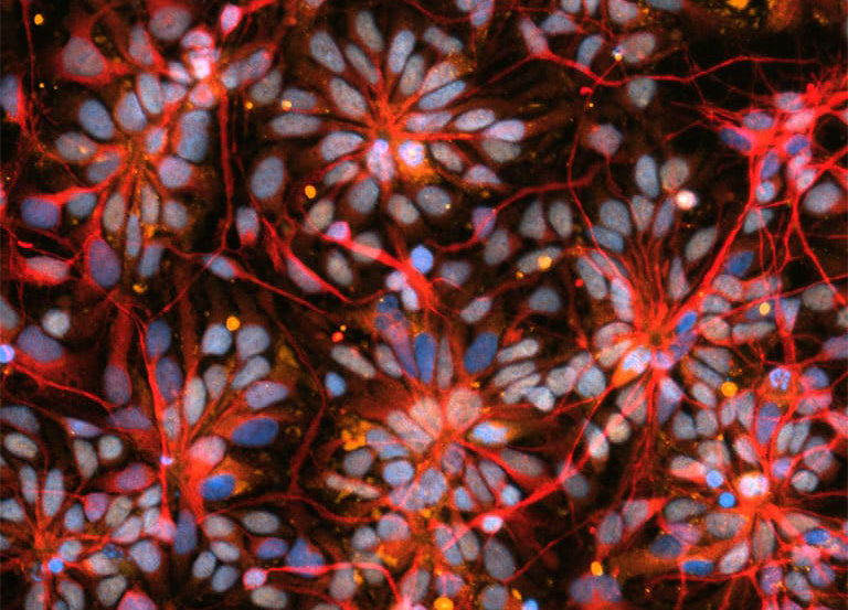
A view of brain organoid cells made of cells from people with Parkinson’s Disease and primary progressive multiple sclerosis.
Media Credit: New York Stem Cell Foundation
Now, with his collaborators at the Federal University of Amazonas in Manaus, Brazil, he is compiling a library of compounds from those plants, asking an AI to predict which molecules might reach the brain, then testing the most promising ones in organoids small enough to fly on a rocket. What used to take years, he says, his students can do in a week. The first of these plant-based compounds will go up early next year.
Saving time was always part of the plan. A brain ages over decades, and by the time anyone notices something is wrong, the damage is often long underway. Up there on the space station, where the signals of that decline run faster for reasons no one can yet explain, researchers may get to watch the unraveling as it happens, and try, finally, to stop it before it starts.